

Clinical Tools
Professional LxH Dynamic Disc Model
Features include:
- new cauda equina with optional rootlets (blue) (2020 update)
- circumferential (diffuse) disc bulge
- superimposed disc protrusion
- limacon shaped annulus
- peripherally exposed calcified endplate
- elastomeric white articular cartilage
- subchondrial bone exposed with a hyaline fibrillation
- bone coloured L5
- white superior endplate matching the colour of the articular cartilage
- blue coloured intervertebral disc innervation
- Flexible and dynamic herniating (or prolapse) nucleus pulposus. This is achieved through a realistic 2-part intervertebral disc with 6 degrees of freedom. Nuclear migration upon manual compression through a torn annulus fibrosus explaining pain generators under load.
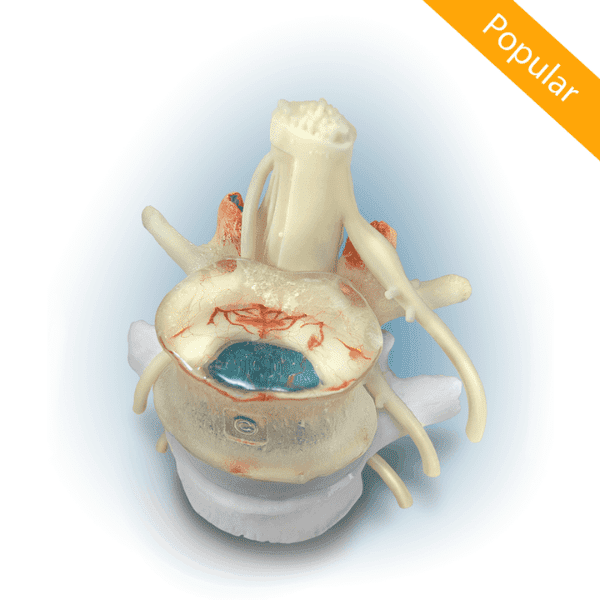
- right posterior-lateral radial and circumferential(concentric) fissure
- intervertebral disc innervation to the outer third of the annulus
- nerve ingrowth (neo-innervation) to the inner two-thirds of the damaged annulus to help demonstrate chronic pain
- left partial posterior-lateral radial tear matching up to the disc protrusion (contained nucleus)
- anterior circumferential tear
- transparent L4
- randomly scattered and embedded black nuclear structures to show nuclear shifting dynamics through the L4 view lens easily
- L5 superior endplate pores (black)
- L5 superior endplate lesion (red)
- vasculature in L4 vertebral body (red)
- facet subchondrial vascularization (red)
- facet tropism (L5 inferior)
- BONUS disc disruption graphic is included as a download
Optional Features: ligamentum flavum (new design), spondylolisthesis (elongated pars, non-lytic)